https://pubmed.ncbi.nlm.nih.gov/41382120/
 https://isid.research.ac.ir/Saeed_Karbasi
 اطلاعیه جلسه دفاعیه دکتری تخصصی - خانم مهندس لیلا جعفری
 https://link.springer.com/article/10.1007/s11481-026-10286-x
 https://www.medrxiv.org/content/10.64898/2026.03.04.26347652v1
 https://journals.lww.com/jmss/fulltext/2026/01020/diagnosing_multiple_sclerosis_from_magnetic.3.aspx
 https://isid.research.ac.ir/Monireh_SheihkHosseini
 https://isid.research.ac.ir/Zahra_Baharlouei
 https://isid.research.ac.ir/Farnaz_Sedighin
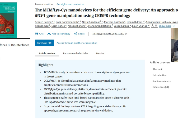 The MCM/Lys-Cys nanodevices for the efficient gene delivery: An approach towards MCP1 gene manipulation using CRISPR technology